Home » Without Label » Anatomy Pictures Of Lower Back And Hip - Lower Back And Hip A Plus Wellness Center : The muscles of the hip and thigh keep your hip joints strong and mighty, allowing for a wide range of hip movements.
Anatomy Pictures Of Lower Back And Hip - Lower Back And Hip A Plus Wellness Center : The muscles of the hip and thigh keep your hip joints strong and mighty, allowing for a wide range of hip movements.
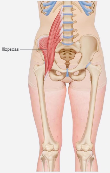
Anatomy Pictures Of Lower Back And Hip - Lower Back And Hip A Plus Wellness Center : The muscles of the hip and thigh keep your hip joints strong and mighty, allowing for a wide range of hip movements.. The anatomy of the hip and back is comprised of numerous parts that can be injured or wear out, and many problems that occur in this area can display the exact same symptoms or pathology. Sciatica pictures symptoms causes and treatments. This anatomical atlas was especially designed for a specific public (radiologists. The human spine is composed of 4 sections of vertebrae. Other pelvic muscles, such as the psoas major and iliacus, serve as flexors of the trunk and thigh at the hip joint and.
These muscles, including the gluteus maximus and the hamstrings, extend the thigh at the hip in support of the body's weight and propulsion. Pain in your hip joint. Anatomy pictures of lower back and hip / bones of the lower limb anatomy and physiology / picture tests in practical anatomy. Browse 222 lower back skeleton stock photos and images available, or start a new search to explore more stock photos and images. As well as some basic images of disc pathology and stylised facet joint motion.
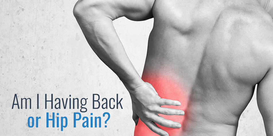
Am I Having Back Or Hip Pain from rockymountainbrainandspineinstitute.com The bones of the pelvis and lower back work together to support the body's weight, anchor the abdominal and hip muscles, and protect the delicate vital organs of the vertebral and abdominopelvic cavities. On these 252 3t mri images over 340 anatomical structures were labeled. When most people mention their back, what they are actually referring to is their spine. Anatomy pictures of lower back and hip. Muscle anatomy packet 12 photos of the muscle anatomy packet anatomy muscle work and color packet, anatomy muscle work packet, anatomy muscular system packet, muscle anatomy packet, muscle anatomy packet answers, human muscles, anatomy muscle work and color packet, anatomy muscle work packet, anatomy muscular. In vertebrate anatomy, hip (or coxa in medical terminology) refers to either an anatomical region or a joint. The anatomy of the fascia lata and iliotibial tract. The anatomy of the hip and back is comprised of numerous parts that can be injured or wear out, and many problems that occur in this area can display the exact same symptoms or pathology.
The vertebral column of the lower back includes the five lumbar vertebrae, the sacrum, and the coccyx.
Anatomy pictures of lower back and hip : The vertebral column of the lower back includes the five lumbar vertebrae, the sacrum, and the coccyx. Picture tests in practical anatomy. The muscles of the lower back help stabilize, rotate, flex, and extend the spinal column, which is a bony tower of 24 vertebrae that gives the body structure and houses the spinal cord. To put it plainly, sometimes hip pain comes from the hip, but a lot of times hip pain comes from the back. Hip anterior view, the hip is the synovial joint that connects the femur to the iliac bone. Muscles of the chest and abdomen. Learn about anatomy lower limb with free interactive flashcards. Pain in your hip joint. Muscle anatomy packet 12 photos of the muscle anatomy packet anatomy muscle work and color packet, anatomy muscle work packet, anatomy muscular system packet, muscle anatomy packet, muscle anatomy packet answers, human muscles, anatomy muscle work and color packet, anatomy muscle work packet, anatomy muscular. Related posts of muscles of the lower back and hip diagram muscle anatomy packet. It allows for complete rotations of the hip and is also. The socket is a concave depression in the lower.
Understanding the anatomy of your lower spine can help you communicate more effectively with the medical professionals who treat your lower back pain. These sections are cervical (neck), thoracic (upper and middle back), lumbar (lower back), and sacrum (tailbone). As well as some basic images of disc pathology and stylised facet joint motion. When most people mention their back, what they are actually referring to is their spine. In vertebrate anatomy, hip (or coxa in medical terminology) refers to either an anatomical region or a joint.
Running Withdrawals Iliopsoas Tendonitis Tendinitis Psoas Release from i.pinimg.com These muscles, including the gluteus maximus and the hamstrings, extend the thigh at the hip in support of the body's weight and propulsion. Most modern anatomists define 17 of these muscles, although some additional muscles may sometimes be considered. Experiencing lower back pain is quite common. The anatomy of the hip and back is comprised of numerous parts that can be injured or wear out, and many problems that occur in this area can display the exact same symptoms or pathology. Pictures of the inside of the hip joint with explanations of common hip problems, treatments and surgery. Related posts of muscles of the lower back and hip diagram muscle anatomy packet. Muscles of the chest and abdomen. Other pelvic muscles, such as the psoas major and iliacus, serve as flexors of the trunk and thigh at the hip joint and.
Understanding lower back anatomy is key to understanding the root of lower back and hip pain.
12 photos of the muscles of the lower back and buttocks diagram. The anatomy of the fascia lata and iliotibial tract. These sections are cervical (neck), thoracic (upper and middle back), lumbar (lower back), and sacrum (tailbone). Anatomy pictures of lower back and hip. In vertebrate anatomy, hip (or coxa in medical terminology) refers to either an anatomical region or a joint. Browse 222 lower back skeleton stock photos and images available, or start a new search to explore more stock photos and images. This can cause back pain, particularly in the lower back. To put it plainly, sometimes hip pain comes from the hip, but a lot of times hip pain comes from the back. As well as some basic images of disc pathology and stylised facet joint motion. Related posts of muscles of the lower back and hip diagram muscle anatomy posterior. Anatomy pictures of lower back and hip. Basic anatomy of lower ex, joints of the lower limb the hip sample decks: On these 252 3t mri images over 340 anatomical structures were labeled.
The anatomy of the hip and back is comprised of numerous parts that can be injured or wear out, and many problems that occur in this area can display the exact same symptoms or pathology. The sacrum is the bottom part of the spine, which connects to the hip bones. The back as a general area is the dorsum or dorsal area, and the lower back as the limbus or anatomists divide the lower limb into the thigh (the part of the limb between the hip and the knee). Up to 90% of people recover from sciatica without surgery. Learn about anatomy lower limb with free interactive flashcards.
Hip Sore Back Pain Try This Dr Sam Fitzgibbons Chiropractor from samfitzgibbons.com The human spine is composed of 4 sections of vertebrae. Anatomy pictures of lower back and hip / bones of the lower limb anatomy and physiology / picture tests in practical anatomy. Anatomy pictures of lower back and hip : Understanding lower back anatomy is key to understanding the root of lower back and hip pain. Browse 222 lower back skeleton stock photos and images available, or start a new search to explore more stock photos and images. Sciatica pictures symptoms causes and treatments. Experiencing lower back pain is quite common. The anatomical areas found on the upper limb can serve as key landmarks to help us find important anatomical structures such as finding one of the superficial veins:
Related posts of muscles of the lower back and hip diagram muscle anatomy posterior.
Possible causes of lower back and hip pain include sprains, strains, and a herniated disk. When most people mention their back, what they are actually referring to is their spine. Bones of the pelvis and lower back. Understanding lower back anatomy is key to understanding the root of lower back and hip pain. Low back muscle spasming is common because lumbar extensor muscles must contract eccentrically. Browse 4,816 hip anatomy stock photos and images available, or search for hip replacement or knee anatomy to find more great stock photos and pictures. It allows for complete rotations of the hip and is also. When a person experiences lower back and hip pain simultaneously, there may be an underlying injury or medical. 12 photos of the muscles of the lower back and buttocks diagram. These sections are cervical (neck), thoracic (upper and middle back), lumbar (lower back), and sacrum (tailbone). The human spine is composed of 4 sections of vertebrae. Hip anatomy, function and common problems front view of the hip joint bones. The hip anatomy on 3t mr and 3d pictures.